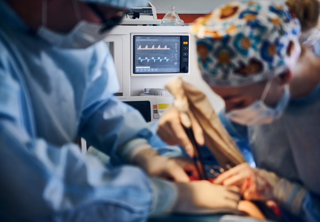
Every year, thousands of people across Malaysia die from coronary artery disease — many without ever knowing they had a problem until it was too late. The frightening reality of blocked arteries is that they rarely announce themselves with obvious symptoms until they are already dangerously advanced. A person can feel perfectly healthy, maintain an active lifestyle, and still be walking around with an artery that is 70 or 80 percent narrowed, silently limiting blood flow to the heart muscle. This is why cardiologists consider the heart catheterization procedure one of the most valuable tools in modern cardiac medicine — not just for treatment, but for life-saving early detection.
Unlike non-invasive imaging tests that provide estimates and probabilities, cardiac catheterization delivers a direct, real-time view of what is happening inside the coronary arteries. It answers the question that every at-risk patient and every concerned cardiologist needs answered with certainty: are there blockages present, how severe are they, and what needs to be done about them?
What Happens Inside an Artery When a Blockage Forms
To understand why the heart catheterization procedure is so effective at detecting blockages, it helps to understand how those blockages develop in the first place. Coronary artery disease is caused by atherosclerosis — the gradual buildup of a fatty, waxy substance called plaque along the inner walls of the arteries that supply blood to the heart. This process begins silently, often in a person’s twenties or thirties, and progresses over decades.
As plaque accumulates, the internal diameter of the artery narrows — a condition called stenosis. In the early stages, the heart compensates, and there are no symptoms. As stenosis progresses past 50 to 60 percent, exertional symptoms like chest tightness and breathlessness may appear. Beyond 70 percent blockage, the restriction of blood flow becomes clinically significant. And at a critical threshold, or when a plaque ruptures and triggers sudden clot formation, a heart attack occurs. The danger lies in the fact that plaque can rupture and cause a heart attack even before a person has experienced any warning symptoms whatsoever.
This unpredictable nature of coronary artery disease is exactly why definitive diagnostic imaging — delivered through cardiac catheterization — matters so much. Stress tests and CT scans can raise suspicion of disease, but catheterization confirms it with a degree of anatomical precision that no other currently available test can match.
How the Heart Catheterization Procedure Maps the Coronary Arteries
The heart catheterization procedure — also known as coronary angiography when used specifically to image the arteries — works by introducing a thin, flexible tube called a catheter into the body’s arterial system and guiding it, under continuous X-ray imaging, to the opening of the coronary arteries. Access is most commonly gained through the radial artery in the wrist, though the femoral artery in the groin is an alternative entry point used in specific clinical situations.
Once the catheter is precisely positioned at the coronary artery opening, the cardiologist injects a small volume of radiopaque contrast dye — a substance that is clearly visible under X-ray fluoroscopy. As the dye flows through the coronary artery system, the imaging equipment captures its movement in real time. Where the artery is open and healthy, the dye flows freely and the artery appears well-defined on the image. Where a blockage or narrowing exists, the dye flow is visibly restricted or interrupted — producing the characteristic appearance that cardiologists use to diagnose, grade, and plan treatment for coronary disease.
The entire imaging sequence captures multiple coronary vessels from multiple angles, producing a comprehensive anatomical map of the heart’s blood supply. Cardiologists assess each segment of each visible artery, quantify the degree of stenosis, identify the location and length of each narrowing, and evaluate the quality of blood flow beyond the blockage. This detailed picture — obtained in a procedure that typically takes 30 to 60 minutes from start to finish — provides the definitive information needed to decide whether a patient needs medical management, a stent procedure, or coronary artery bypass surgery.
Why Early Detection Through Cardiac Catheterization Saves Lives
The clinical case for early detection of coronary blockages is compelling and extensively supported by cardiac outcome research. A patient whose 75 percent coronary stenosis is identified through a planned diagnostic procedure — and treated with a coronary stent before a heart attack occurs — faces a fundamentally different prognosis than a patient whose first presentation of the same disease is an acute myocardial infarction requiring emergency intervention.
Elective, planned cardiac catheterization performed in a fully equipped facility allows the cardiologist to assess the anatomy calmly and completely, plan the intervention with precision, and execute it under optimal conditions. Emergency catheterization performed on a patient in cardiogenic shock — where every minute of delay translates into additional cardiac muscle death — is a higher-risk, more technically demanding procedure with less predictable outcomes, regardless of the facility or the operator’s skill.
Beyond the immediate intervention, early identification of coronary disease allows patients and their care teams to implement aggressive medical management: antiplatelet therapy to reduce clotting risk, lipid-lowering treatment to stabilise existing plaque, antihypertensive management, and structured cardiac rehabilitation. These secondary prevention measures, applied after a known diagnosis but before a catastrophic event, dramatically reduce the risk of future cardiac events. They cannot be applied to a disease that has not yet been diagnosed.
Who Should Consider Discussing Cardiac Catheterization With Their Doctor
Not every person needs or should have a heart catheterization procedure — it is an invasive investigation recommended when clinical judgment and less invasive testing indicate a meaningful probability of significant coronary disease. However, certain presentations and risk profiles make a conversation with a cardiologist about catheterization not just reasonable but important.
Patients who should discuss this with their doctor include those who experience chest pain or pressure during exertion that is not fully explained by non-cardiac causes; those who have had a positive or equivocal stress test indicating possible ischaemia; those with established risk factors — diabetes, hypertension, high cholesterol, smoking, a strong family history of early coronary disease — who develop new or changing cardiac symptoms; and those who have already had a heart attack and require anatomical assessment before discharge or treatment planning.
Asymptomatic patients in high-risk categories may also be candidates when non-invasive imaging raises sufficient concern. The specific threshold for recommending catheterization reflects a careful weighing of the diagnostic benefit against the small but real risks of the procedure — a conversation that only a qualified cardiologist, with access to the patient’s full clinical picture, can properly conduct.
What Patients Can Expect: Before, During, and After
Preparation for a heart catheterization procedure typically involves fasting for several hours before the procedure, withholding certain regular medications as directed by the care team, and providing a detailed medication and allergy history — particularly regarding iodine-based contrast agents and blood thinners. Blood tests and an electrocardiogram are usually obtained beforehand.
During the procedure, patients are awake but sedated — comfortable and relaxed but able to respond to the cardiologist’s instructions, such as holding a breath briefly during image acquisition. The access site is numbed with local anaesthetic, and the catheter insertion causes minimal sensation. Patients often feel a brief warmth or flushing when the contrast dye is injected — a normal and temporary sensation. The procedure room is staffed by a cardiologist, a cardiac nurse specialist, and a radiographer or technologist operating the imaging equipment.
Recovery for radial (wrist) access cases is typically brief — a few hours of observation with a compression device applied to the access site, followed by discharge home on the same day for elective procedures. Activity restrictions are minor and short-lived compared to femoral access, which has contributed to radial access becoming the preferred approach in most contemporary catheterization laboratories.
Where to Access Heart Catheterization Services in Malaysia
For patients in Melaka and the surrounding region seeking expert cardiac investigation, Putra Hospital Melaka provides dedicated cardiac catheterisation laboratory services staffed by experienced interventional cardiologists supported by purpose-built, modern imaging infrastructure. The facility is equipped to perform both diagnostic coronary angiography and therapeutic interventional procedures — including coronary stenting — within the same setting, allowing patients to move from diagnosis to treatment without the delays and transfer risks associated with facilities that offer diagnosis only.
Access to a dedicated catheterization lab with experienced cardiac imaging specialists matters significantly for procedure quality and patient safety. Advanced flat-panel detector fluoroscopy systems deliver high-resolution arterial imaging at reduced radiation doses compared to older equipment. Experienced teams who perform catheterization regularly develop the procedural fluency and case familiarity that translate directly into better patient outcomes and lower complication rates.
Patients who have been referred by their cardiologist for further investigation, or who have risk factors and symptoms that have not yet been fully evaluated, are encouraged to discuss the heart catheterization procedure with a specialist rather than allowing uncertainty about the state of their coronary arteries to persist. The information a catheterization delivers — whether it confirms disease that needs treatment or provides reassurance that the arteries are clear — is uniquely valuable information that no other investigation currently provides with the same clarity and precision.
Frequently Asked Questions
Q: Is the heart catheterization procedure painful?
A: Most patients report that the procedure is significantly more comfortable than they anticipated. The access site — usually the wrist — is numbed with local anaesthetic before catheter insertion, and sedation keeps patients relaxed throughout. A brief warmth or flushing sensation when contrast dye is injected is normal and temporary. Significant pain during the procedure is uncommon and should be reported immediately to the cardiac team.
Q: How accurate is the heart catheterization procedure at detecting blockages?
A: Coronary angiography performed during cardiac catheterization is considered the reference standard diagnostic test for coronary artery disease. Its direct visualisation of contrast flow through the arterial lumen provides anatomical detail and stenosis quantification that non-invasive imaging approaches cannot match. It identifies blockages, their precise location, their severity, and the extent of the coronary territory at risk — information essential for treatment planning.
Q: What is the difference between a diagnostic catheterization and an interventional one?
A: A diagnostic catheterization — coronary angiography — is performed solely to visualise the arteries and identify any blockages present. An interventional catheterization proceeds further: after the diagnostic images identify a significant blockage, the cardiologist uses the same access to advance specialised equipment to the blockage site and performs a treatment such as balloon angioplasty or coronary stenting. Many centres, including facilities like Putra Hospital Melaka, are equipped to perform both in a single session when the clinical situation is appropriate.
Q: How long does recovery take after a heart catheterization procedure?
A: Recovery following an elective diagnostic procedure performed through radial (wrist) access is typically four to six hours of observation before discharge. Patients can usually return to light daily activities within 24 hours and normal activity within two to three days. The care team will provide specific activity guidelines and instructions regarding the access site, medications, and any signs that should prompt urgent review.
Q: Can the procedure detect blockages that are not yet causing symptoms?
A: Yes, and this is one of its most important clinical applications. Significant coronary stenosis — narrowings that restrict blood flow enough to put the patient at elevated heart attack risk — can exist without causing any noticeable symptoms in daily life. The heart catheterization procedure identifies these anatomically significant blockages regardless of whether they are producing symptoms, enabling treatment and secondary prevention to be initiated before a potentially fatal cardiac event occurs.